Treatment of uterine fibroids
Uterine fibroids are benign (non-cancerous) tumors that develop on the surface or inside uterine muscle tissue
Uterine fibroids

What is a fibroid?
Uterine fibroids are benign tumours (non-cancerous) that develop on the surface or inside the uterine muscle tissue.
In many women, the presence of uterine fibroids goes completely unnoticed. In others, fibroids location and size can affect the quality of life.
What discomfort can fibroids cause?
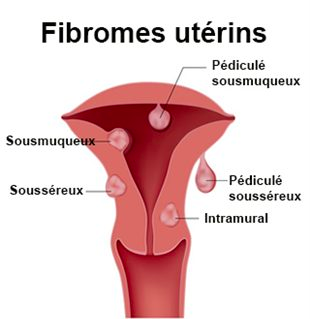
Depending on the case, there can be multiple symptoms or non-at all. It all depends on the size and location of the fibroid (see diagram). Fibroids are sensitive to hormones; their symptoms are often related to the menstrual cycle. Just before menopause when the oestrogen level tends to increase, their size tends to grow, resulting in an elevation of the symptoms. Once the menopause set in, oestrogen levels decrease significantly. Fibroids and their symptoms are reduced accordingly.
The most common symptoms are:
Depending on their size and location, fibroids can also interfere with the implantation and development of a pregnancy.
What are the existing medical treatments?
Many doctors use the birth control pill with progestin to control excessive menstrual bleeding caused by fibroids. The doctor may also prescribe non-steroidal anti-inflammatory drugs for pain relief. The GnRH agonists have the effect of reducing the production of oestrogen in the ovaries. They help reduce the size of fibroids and limit the symptoms. Due to the oestrogen level decrease, there are side effects such as hot flashes or mood swings. This type of product is thus difficult to use over a long period.
What are the surgical options?
The ablation of fibroids, operation called “myomectomy,” can be achieved through different techniques:
What is the embolization place?
The uterine fibroid embolization is a medical procedure developed in France in the early 90’s. The principle of embolization is to deprive fibroids of food and blood through the injection of synthetic microbeads in the uterine arteries. This technique has its place as an alternative to surgery. However, all patients cannot benefit from it and the embolization decision must be made on a case-by-case basis.
Once the choice of embolization has been made, a team involving the interventional vascular radiologist, the gynaecologist and the anaesthetist is formed.
The gynaecologist establishes a medical assessment based on the information obtained during the imaging tests (ultrasound, MRI); the interventional vascular radiologist makes sure there is no contraindication and plans the embolization procedure; the anaesthetist sets the associated medical treatment (pain killer).
An appointment with the interventional radiologist is organized to explain the course of embolization and answer questions that the patient may have.
The procedure is performed under local anaesthesia supplemented by sedation or also under loco-regional anaesthesia. This procedure is done by puncturing the femoral artery and placing of a catheter through which the radiologist will inject microbeads which have the effect of depriving the fibroid from its vascularity. The process takes 24 to 72 hours of hospitalization with a leave of absence from work of about 10 days.
There can be side effects and failures.


